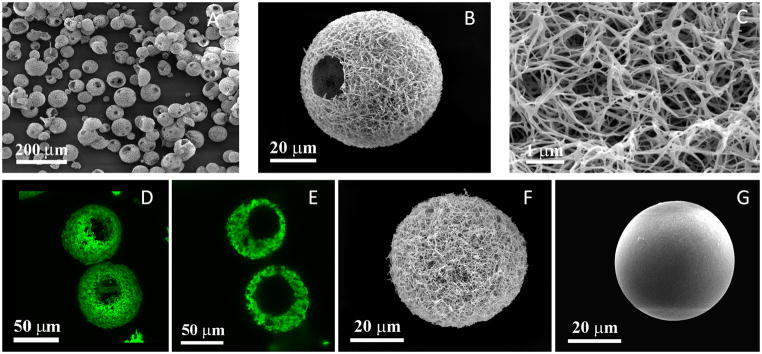
Figure 2. Characterization of nanofibrous hollow microspheres, nanofibrous microspheres, and solid interior microspheres.
(A) SEM image of nanofibrous hollow microspheres fabricated from SS-PLLA, noting that almost every microsphere had one or more open hole(s) on the shell. (B) SEM image of a representative nanofibrous hollow microspheres, showing the nanofibrous architecture and a hole of approximately 20 μm on the microsphere shell. (C) A high magnification image of (B) showing the nanofibres of the microsphere with an average diameter of about 160 nm. (D) A 3D reconstruction of nanofibrous hollow microspheres from confocal image stacks. (E) A 2D cross-section confocal image of the nanofibrous hollow microspheres, confirming the hollow structure of the microsphere. (F) SEM image of a representative nanofibrous microsphere, showing a nanofibrous architecture on the microsphere surface. (G) SEM image of a representative solid interior microspheres, showing a smooth surface of the microsphere. All the microspheres shown in this figure were fabricated from a 2% (wt/v) polymer solution. The nanofibrous hollow microspheres were fabricated from SS-PLLA. Both nanofibrous microspheres and solid interior microspheres were fabricated from a linear PLLA with an inherent viscosity of approximately 1.6 dl/g.

