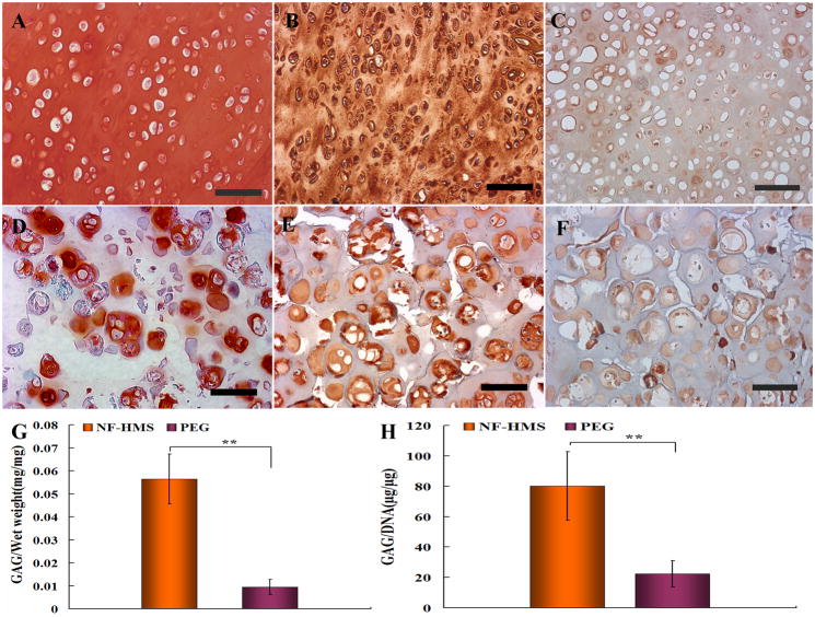
Figure 5. Comparison between neo cartilage tissues regenerated from nanofibrous hollow microspheres/chondrocytes and PEG/chondrocytes constructs after 8 weeks of subcutaneous implantation in nude mice.
Histological examination showed that neo-cartilage tissue regenerated from the nanofibrous hollow microspheres/chondrocytes group contained uniform GAG-rich ECM as evidenced by safranin-O staining (A), and abundant collagen type II as shown by immunohistochemical staining (B). In contrast, only pericellular regions of the chondrocytes were stained positively for GAG (D) and collagen type II (E) in the PEG/chondrocytes group. While the collagen type II was significantly more abundant in the nanofibrous hollow microspheres/chondrocytes group than in the PEG/chondrocytes group, the collagen type I staining was similarly weak in overall intensity between the two groups (C and F). Biochemical analysis (n=6) revealed that cartilage tissue formed from the nanofibrous hollow microspheres (NF-HMS)/chondrocytes constructs had significantly higher GAG/wet weight ratio (G) and GAG/DNA ratio (H) than those from the PEG/chondrocytes constructs. Error bars in panels G & H indicate standard deviation. ** p<0.01.

