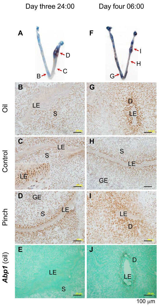
Figure 4.
Immnuostaining of progesterone receptor (PR) and in situ hybridization (ISH) detection of decidualization marker amiloride-binding protein 1 (Abp1) in mouse uterus with artificial decidualization. Longitudinal frozen sections (10 µm) crossing luminal epithelium were used. A. A representative uterine image on day three 24:00 hours: left uterine horn with oil injection, right uterine horn with two pinches near the uterotubal junction, red arrows indicating the areas sectioned for PR immunohistochemistry (IHC) in B~D. B. PR IHC, day three 24:00 hours, oil-injected uterine horn. C. PR IHC, day three 24:00 hours, control uterine segment without stimulation; an enlarged view of the white boxed area shown in the insert on left bottom corner; the seemingly multiple luminal epithelial (LE) layers indicating a particular sectioning angle that cut through several LE cells on the single LE layer. D. PR IHC, day three 24:00 hours, a pinched area. E. Abp1, ISH, antisense probe, day three 24:00 hours, the same oil injected area as in B. Abp1 is undetectable at this hour. F. A representative uterine image on day four 06:00 hours: left uterine horn with oil injection, right uterine horn with two pinches near the uterotubal junction, red arrows indicating the areas sectioned for PR IHC in G~I. G. PR IHC, day four 06:00 hours, oil-injected uterine horn. H. PR IHC, day four 06:00 hours, control uterine segment without stimulation. I. PR IHC, day four 6:00 hours, a pinched area. J. Abp1, ISH, antisense probe, day four 06:00 hours, the same oil injected area as in G. Abp1 is detectable in the subepithelial stromal area at this hour. LE, luminal epithelium; S, stroma; GE, glandular epithelium; D, decidual zone; scale bar, 100 µm. N=3. No specific staining was detected in the negative control for PR IHC or using a sense probe for Abp1 ISH (data not shown).

