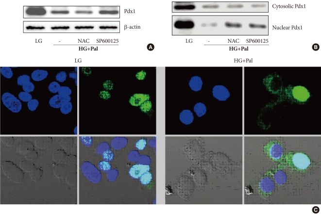
Fig. 4.
Pancreatic and duodenal homeobox 1 (Pdx1) protein expression and nucleo-cytoplasmic translocation of Pdx1 in INS-1 cells after treatment with 30 mM glucose and 0.25 mM palmitate. (A, B) Total proteins and nuclear proteins of Pdx1 were analyzed using Western blotting. (C) The staining of Pdx1 was performed as described in Materials and Methods. The cells were stained with anti-GFP antibody (1:100, green) to detect Pdx1 and with DAPI (blue) to detect nuclei. LG, 5.6 mM glucose; HG, 30 mM glucose; Pal, 0.25 mM palmitate; NAC, N-acetyl-L-cysteine.

