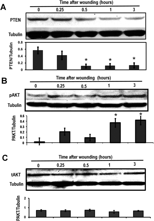
Figure 1.
PTEN was downregulated and Akt was activated after wounding of the HCE cell monolayer, as determined by Western blot analysis. (A) PTEN was increasingly downregulated in a time-dependent manner after wounding. Time 0 = unwounded. (B) pAkt levels were upregulated in a time-dependent manner. The results of Western blot analysis showed that pAkt increased over time. (C) Total Akt levels showed no variation in nonwounded or wounded HCE monolayers. Proteins were isolated using RIPA buffer at 0.25, 0.5, 1, and 3 hours after wounding/without wound. Tubulin was used as a protein loading control. Scanning densitometry of Western blot analysis under each western image was from three different experiments. Bars show mean ± SD. *P < 0.05 compared with unwounded control (T0).

