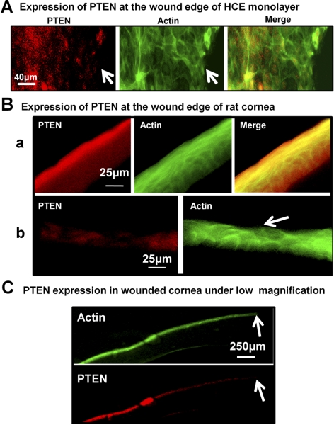
Figure 2.
PTEN was downregulated around wound edges in the HCE cell monolayer and rat cornea. (A) PTEN expression was reduced near the wound edges in the HCE cell monolayer at 1 hour after wounding. (B) In the normal cornea, (a) PTEN expression in the epithelium was higher on the surface than on the lower epithelium of the cornea. After wounding of the cornea, (b) PTEN expression was reduced near the wounded epithelium at 1 hour after wounding, when partial scraping of corneal epithelium, as indicated by the arrow. (C) Under low magnification, PTEN was downregulated distally from the wound edge in the cornea with the full thickness of the epithelium wound. All samples were collected at 1 hour after wounding. Red: PTEN expression was determined by immunofluorescence; green: actin expression; arrow: wound edge.

