Figure 1.
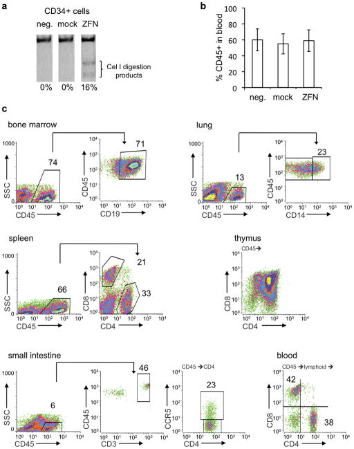
ZFN-mediated disruption of CCR5 gene in HSC. (a) Representative gel showing extent of CCR5 disruption in CD34+ HSC, 24 hours after cells were nucleofected with ZFN-expressing plasmids (ZFN) or mock nucleofected (mock). Neg. is untreated HSC. CCR5 disruption was measured by PCR amplification across the ZFN target site, followed by Cel I nuclease digestion and quantitation of products by PAGE. (b) Graph showing mean +/− SD percentage of human CD45+ cells in peripheral blood of mice at 8 weeks post-transplantation with either untreated, mock nucleofected, or ZFN nucleofected HSC, (n=5 each group). (c) FACS profiles of human cells from various organs of one representative mouse transplanted with ZFN-treated HSC. Cells were gated on FSC/SSC to remove debris. Staining for human CD45, a pan leukocyte marker, was used to reveal the level of human cell engraftment in each organ. CD45+ gated populations were further analyzed for subsets, as indicated: CD19 (B cells) in bone marrow, CD14 (monocytes/macrophages) in lung, CD4 and CD8 (T cells) in thymus and spleen, and CD3 (T cells) in the small intestine (lamina propria). The CD45+ population from the small intestine was further analyzed for CD4 and CCR5 expression. Peripheral blood cells from CD45+ and lymphoid gates were analyzed for CD4 and CD8 expression. The percentage of cells in each indicated area is shown. No staining was observed with isotype-matched control antibodies (Supplementary Fig. 1 online), or in animals receiving no human graft (data not shown).

