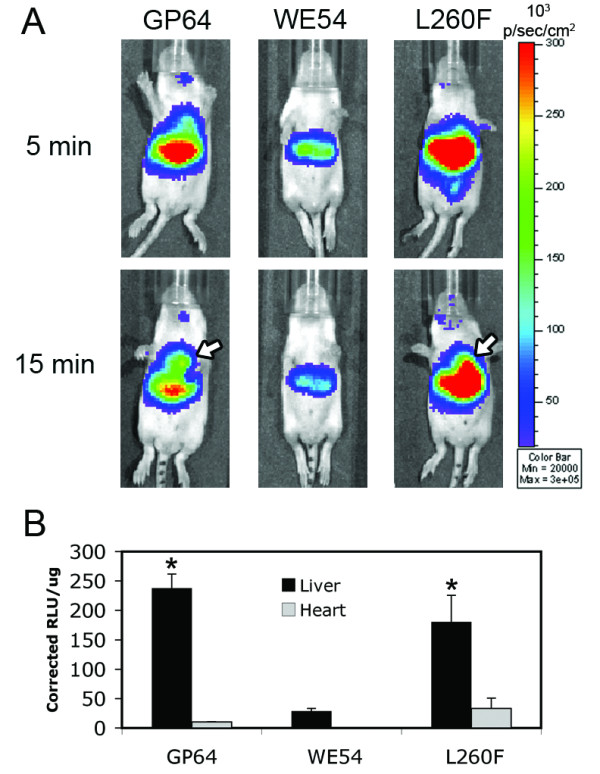
Figure 5.

Neonatal delivery of FIV-L260F transduces the liver and heart. One week after facial vein injection of pseudovirions, mice were imaged utilizing a CCD camera. (A) Representative photos of mice 1 week postinjection are depicted following injection with FIV-GP64, FIV-WE54, and FIV-L260F. Waiting an additional 10 minutes after IP delivery of D-luciferin allowed for luciferase expression to be detected from the hearts (arrows) in animals transduced with FIV-GP64 and FIV-L260F, while the hearts of FIV-WE54 transduced animals displayed no detectable signal. (B) Three weeks postinjection, the heart, lungs, liver, spleen, and kidneys were harvested and luciferase assays conducted. Expression was detected exclusively from liver tissue (black bars) for all pseudoviruses tested and from hearts (gray bars) for the GP64 and L260F pseudoviruses. Standard errors are plotted and * denotes statistical significance (P ≤ 0.05) compared to FIV-WE54 livers. n = 5 for FIV-GP64 and FIV-WE54, 8 for FIV-L260F.
