Figure 5.
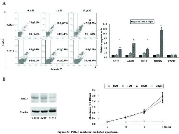
PRL-3 inhibitor-mediated apoptosis. (A) Apoptosis assay was performed 72 hours after treatment with PRL-3 inhibitor (0 to 10 μmol/L). Representative figures of apoptosis assay on AZ521 and C2C12 cells were shown in left panel, and the percentage and SD of early apoptosis (bottom right quadrant) and late apoptosis (top right quadrant) are shown in each panel. With cells treated with chemical solution alone (0 μmol/L PRL-3 inhibitor) as 1.0, the relative late apoptosis rate after treatment was shown in right panel. *, P < 0.05 by Student t test, compared with 0 μmol/L of PRL-3 inhibitor. (B) PRL-3 inhibitor treatment against normal skeletal muscle C2C12 cells. C2C12 cells exhibited lower expression level of PRL-3 than GC cells by western blotting. Proliferation assay after treatment with PRL-3 inhibitor was performed. Error bars, SD.

