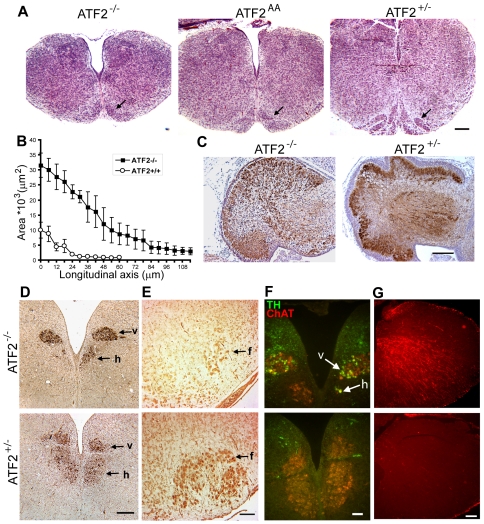
Figure 1. Histological abnormalities in E18.5 ATF2 mutant cerebellum and brainstem.
(A) Hematoxylin and eosin (H&E) stained transversal sections of brainstem at the level of the inferior olive. Atf2−/− and Atf2AA brainstems are significantly smaller and have an enlarged central canal compared to Atf2+/−. The inferior olive (arrow) is severely underdeveloped in mutant embryos. Bar: 250 µm. (B) Area plot of the central canal (mean ± SEM of 3 brainstems analysed per genotype), measured along the antero-posterior axis of serial transversal sections from the obex (0 µm on the x-axis) posteriorly to the caudal end of the medulla, shows a significant enlargement of the canal in Atf2−/− embryos. (C) Horseradish peroxidase (HRP) immunostaining of calbindin in sagittal sections of the cerebellum. Atf2−/− cerebellum lacks the foliation and the tripartite layering seen in Atf2+/− cerebellum. Bar: 250 µm. (D) HRP immunostaining of choline acetyltransferase (ChAT) in transversal sections of Atf2−/− and Atf2+/− posterior medulla. Number of hypoglossal motoneurons (h) is bilaterally decreased in Atf2−/− embryos while number of dorsal vagal motoneurons (v) appears normal. This was seen at several levels along the longitudinal axis. Bar: 200 µm. (E) HRP immunostaining of Islet-1 (Isl-1) in transversal sections of Atf2−/− and Atf2+/− anterior medulla. A severe reduction in the number of motoneurons is found in the Atf2−/− facial nucleus (f). Bar: 100 µm. (F) Double immunofluorescence staining of TH (green) and ChAT (red) shows aberrant expression of TH in hypoglossal (h) and dorsal vagal (v) motoneurons in Atf2−/− brains. Bar: 50 µm. (G) GFAP immunostaining (red) reveals aberrant expression of GFAP in the mantle zone of Atf2−/− brainstem. Bar: 50 µm.

