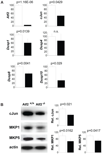
Figure 8. Expression of ATF2 target genes in hindbrain motoneurons.
Real-time quantitative PCR assays of laser micro-dissected tissue of E14.5 hindbrains. Bar graphs show relative expression values between Atf2+/+ (white bars) and Atf2−/− (black bars) for Atf2, c-Jun, Dusp1, Dusp4, Dusp8, and Dusp10 mRNAs. Significance values (p) were determined from 3 independent samples using Student's t-test.

