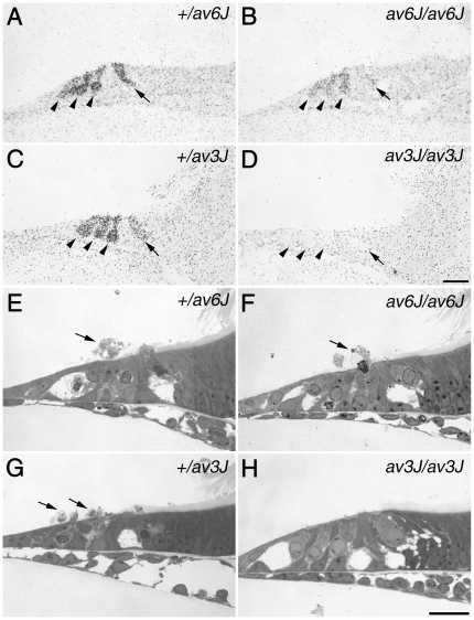
Figure 5. [3H]-gentamicin labelling and gentamicin toxicity in av6J and av3J cochlear cultures.
(A–D) Autoradiographs of [3H]-gentamicin uptake in +/av6J (A), av6J/av6J (B), +/av3J (C) and av3J/av3J (D) apical-coil hair cells. Arrowheads indicate outer hair cells, arrows indicate inner hair cells. [3H]-gentamicin uptake observed in the av6J/av6J mouse (B) is reduced relative to that in the +/av6J control (A). In contrast, av3J/av3J hair cells (D) completely fail to load with [3H]-gentamicin whilst +/av3J control loading (C) is similar to that observed for +/av6J hair cells (A). (E–H) Toluidine blue stained light micrographs of gentamicin treated cochlear cultures from +/av6J (E), av6J/av6J (F), +/av3J (G) and av3J/av3J (H) mice. Arrows indicate extruded hair cells. Whilst +/av6J (E), av6J/av6J (F) and +/av3J hair cells (G) are all sensitive to gentamicin, av3J/av3J hair cells (H) are resistant to this antibiotic. Scale bars = 20 µm.

