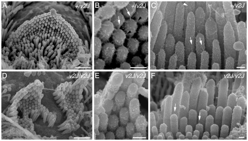
Figure 10. Tip link morphology in v2J cochlear hair cells.
Links from +/v2J (A–C) and v2J/v2J (D–F) mice at P4. (A) Heterozygous +/v2J OHC row 1 hair bundle. The normal row structure is evident at this stage and the stereocilia are well organised. (B) Detail from the bundle shown in A. Normal tip links (white arrows) and lateral links (black arrowheads) are evident near the tips of the stereocilia. (C) Heterozygous +/v2J IHC bundle showing well defined tip links (white arrows), including some that are forked (white arrowhead). (D) Hair bundle from a homozygous v2J/v2J apical row 1 OHC. The bundle is distorted by a central split and there is unevenness in the height of the stereocilia within a row, especially the tallest row. (E) Detail of the bundle shown in D. There is little evidence of robust, full-length tip links (compare B and E). (F) IHC from a v2J/v2J mouse. Occasional stumpy links are evident near the tips of the stereocilia (white arrows). Scale bars = 1 µm in A and D; 200 nm in B, C, E and F.

