Fig. 1.
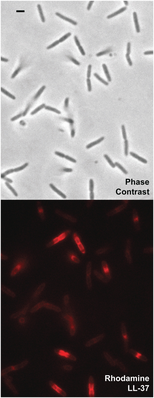
Preferential phase 2 attack on septating cells at t = 12 min after exposure to 8 μM Rh-LL-37. The phase contrast image (Upper) shows cells at different phases of the cell cycle; septating cells look pinched at the midplane. The red fluorescence image (Lower) shows bright bands of Rh-LL-37 centered on the midplane of septating cells (phase 2 attack). Nonseptating cells exhibit only a dim, uniform coating of Rh-LL-37 (phase 1 attack). (Scale bar, 2 μm.)

