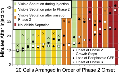
Fig. 3.
The timeline of events for 20 cells in a single experiment. Cells contained periplasmic GFP and were attacked by 8 μM Rh-LL-37. There was no Sytox Green present. The cells were split into four categories based on when septation was visible by eye in the phase contrast channel. Septation likely begins before the time at which it is observable. Phase 2 begins later for cells that are not septating at the beginning of the experiment. In most cases, phase 2 seems to be enabled by the onset of septation. In a few cases for which visible septation is never observed, phase 2 still occurs but is more diffuse in space (Fig. S3). In cases where an event is not shown for a given cell, that event was not analyzable.

