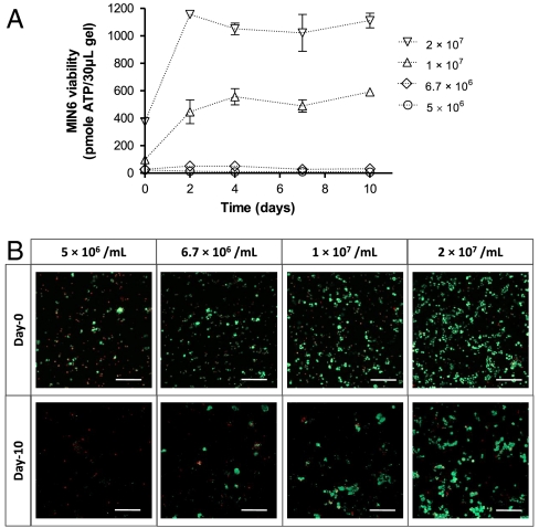
Fig. 1.
Effects of cell-packing density on the survival of MIN6 cells in PEG hydrogels. MIN6 cells were dispersed into single cells and encapsulated at different cell densities as indicated. Cell-laden hydrogels were maintained in RPMI-1640 medium containing 1% FBS. (A) Intracellular ATP of encapsulated MIN6 cells was measured at the indicated time after encapsulation and was used to represent cell metabolic activity and viability (Mean ± SEM, n = 3). (B) Representative confocal Z-stack (300 μm) images of encapsulated MIN6 cells stained with a live/dead viability staining kit at day-0 and day-10. Live cells were stained green while dead cells were stained red (scale: 200 μM).

