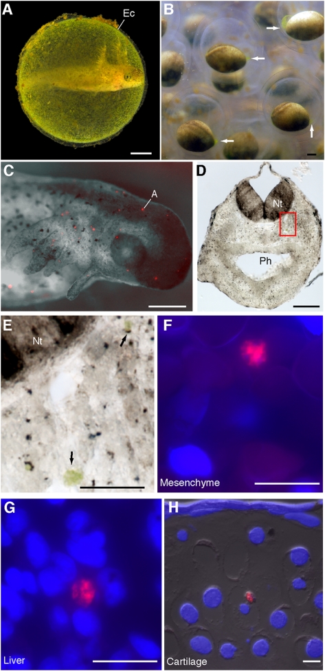
Fig. 1.
Algal cells from the egg capsule invade salamander tissues. (A) A Stage-44 embryo inside its egg capsule (Ec). (B) Still frame taken from time-lapse recordings of Stage-15 embryos. Concentrations of algae occur synchronously, adjacent to the blastopore (arrows). (C) Black-and-white and fluorescent overlay of a Stage-39 embryo removed from its egg capsule; lateral view of the head. Red spots are autofluorescing cells (A) described in text. (D and E) Scattered algal cells embedded in Stage-37 cranial mesenchyme appear green under light microscopy. Boxed region in D shown magnified in E. (F–H) FISH-stained algal cells embedded in (F) Stage-37 cranial mesenchyme, (G) Stage-46 liver, and (H) Stage-46 ceratohyal chondrocyte. Nt, neural tube; Ph, pharynx. (Scale bars: 1 mm in A–D; 50 μm in E; 20 μm in F–H.)

