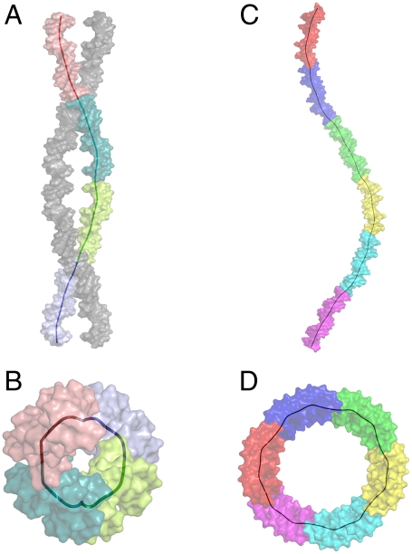
Fig. 5.
Superhelical DNA trajectories in the crystals. The trajectory of the global axes of the colored coil depicts the superhelix. DNA molecules are represented as surface models. Each color of the multicolored DNA coil corresponds to a full response element. A and B show the 10-bp DNA packing perpendicularly and down the DNA left-handed superhelical axis, respectively (A shows the two interwoven coils). There is a quarter-superhelical turn per 20-bp DNA. C and D show the 22-bp DNA perpendicularly and down the right-handed superhelix. There is a one-sixth superhelical turn per 22-bp DNA.

