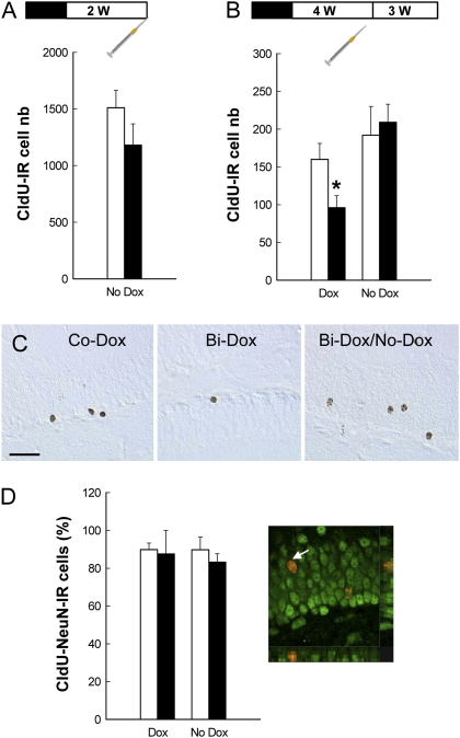
Fig. 3.
Effect of Dox treatment and Dox removal on cell genesis, survival, and differentiation. All mice were first treated for 4 wk with Dox (black boxes in the experimental design). Then Dox was removed (white boxes), and the mice were treated with the sole vehicle for 2 wk (A) or 4 wk (B and D). The syringes represent CldU injections. (A) At 2 wk after Dox removal, cell genesis returned to control values (Co-Dox, n = 5; Bi-Dox, n = 6; t9 = 1.369; P = 0.20). (B) Dox was removed for one subgroup of animals from each genotype for 4 wk (Co-Dox, n = 10; Bi-Dox, n = 9). Dox treatment decreased the number of 3-wk-old CldU-IR cells in bigenic mice (t18 = 2.34; P = 0.03), an effect abolished when Dox was removed from the drinking solution (t17 = −0.35; P = 0.72). (C) Illustration of CldU-IR cells in a Co-Dox mouse, a Bi-Dox mouse, and a Bi-Dox/No-Dox mouse. (Scale bar: 50 μm.) (D) Cell differentiation measured by the percentage of CldU-IR cells expressing the mature neuronal marker NeuN was similar among the groups. (Right) A CldU-IR cell (red stain) expressing NeuN (green stain).

