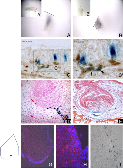
Fig. 2.
Pericytes can respond to injury locally, are involved in tooth development, and are slow-cycling cells. By culturing the X-LacZ4 incisors following damage, and thus eliminating the circulatory system, an increase in the number of LacZ-positive pericytes was evident in both pulp body (A) and cervical loop parts (B), compared with the control incisors (A′ and B′). These cells were characteristically dispersed throughout the pulp tissue, losing their organization within the main body plexus in the pulp body (A and A′), and were intensely concentrated on the bottom edge of the cervical loop area (B and B′), where a vessel-rich area is present. Coexpression of proliferation marker PH3 and LacZ-positive (NG2-positive) pericytes following in vivo damage (C and C′) showed that proliferating pericytes (black arrows) are located on blood vessels extending toward the damaged area (C′). During tooth development (bud to bell stages) in X-LacZ4 mice, LacZ-positive cells are found under the condensing mesenchyme (D) and in the blood vessels close to the forming tooth (E). When colocation of migrating slow-cycling cells and pericytes were analyzed by transplantation experiments associated with nucleoside (i.e., IdU) administration, some cells that migrated from the X-LacZ4 host were both LacZ-positive (pericytes, black stained cells) and IdU positive (slow-cycling, red stained cells) (G and H). These cells were found in the blood vessel-rich cervical loop area of incisors (F and G). Consecutive sections showed that these cells were rare but clustered (G), suggesting a spatial organization. High magnification showed colocalization of IdU and LacZ-positive pericytes under fluorescence (H) and bright field (I).

