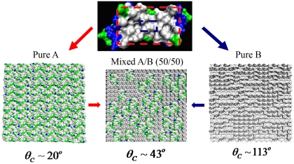
Fig. 3.
Melittin-based surfaces comprising protein fragments of chosen type: (Upper) 2MLT (melittin dimer): The part inside the red rectangle represents a patch of type A, and the part inside blue rectangle represents a patch of type B. (Lower Left) Randomized hydrophilic surface comprising flattened and replicated patches of type A. (Lower Right) Randomized hydrophobic surface prepared by replicating patches of type B. (Lower Center) Randomized mixed A/B (50/50) surface prepared by mixing patches of type A and equal-size domains of six smaller patches B. Gray color represents hydrophobic residues; other colors represent hydrophilic residues. Among the latter, green color denotes neutral and hydrophilic and blue denotes basic and hydrophilic.

