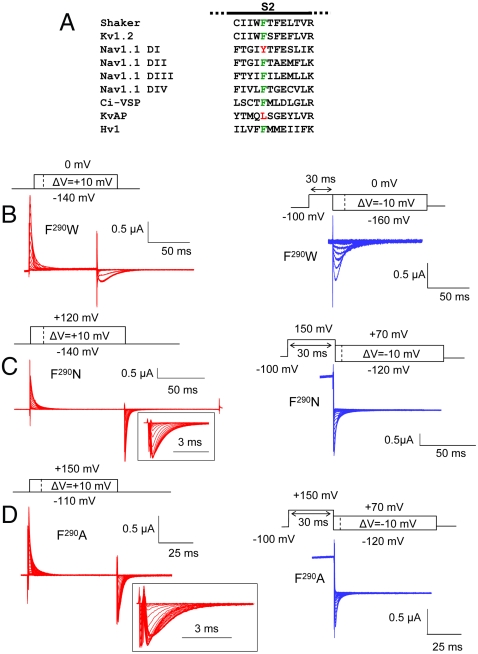
Fig. 1.
(A) Sequence alignment of the S2 segment of Shaker (GI: 13432103), Kv1.2 (GI: 4826782), Nav1.1 domain I, II, III, and IV (GI: 115583677), Ci-VSP (GI: 76253898), KvAP (GI: 38605092), and Hv1 (GI: 91992155). The residue at position 290 (in Shaker) is colored (green for Phe, red for other amino acid). (B–D) Gating currents of the representative mutants F290W (B), F290N (C), and F290A (D) during the indicated activation (red traces) or deactivation (blue traces) protocol. Capacitive and leak currents were subtracted either online using the P/4 method (F290W) or using an off-line subtraction (F290N and F290A). Gating current traces at the repolarizing pulses of the activation protocol are shown in an expanded time scale for the mutants F290N and F290A (insets).

