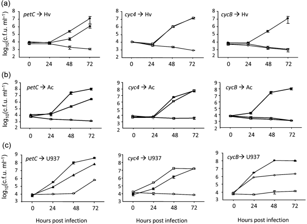
Fig. 6.
Intracellular infection by L. pneumophila wild-type and c-type cytochrome mutants. H. vermiformis (Hv, a), A. castellanii (Ac, b), and U937 cell macrophages (c) were infected with wild-type 130b (•) and ccmC mutant NU295 (○) (all panels in a–c) as well as the petC mutant NU375 (▴) (left panels), cyc4 mutant NU379 (□) (centre panels), and cycB mutant NU381 (◊) (right panels), and then at the indicated times, the c.f.u. in the infected wells were determined by plating. Data are the mean and standard deviations from four infected wells (error bars not shown where smaller than symbols). In (a) and (b), the recovery of the petC and cycB mutants was significantly less than that of wild-type at 48 and 72 h (Student's t-test, P<0.05). In (c), the recovery of the petC and cycB mutants was less than that of wild-type at 24, 48, and 72 h (Student's t-test, P<0.05). Each of the cytochrome mutants was tested on at least two occasions, with the results obtained being comparable to those depicted here. Although not shown here for the sake of space, additional experiments using NU376 and NU382 confirmed the infectivity defects of the petC mutant and cycB mutant.

