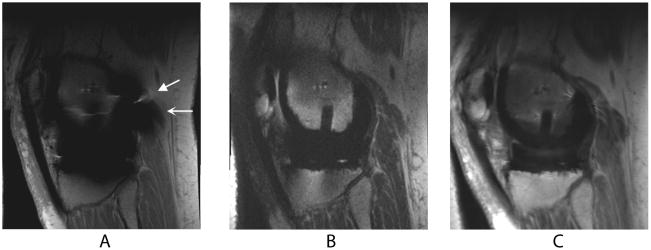
Figure 1.
Sagittal MR images of a TKR in a healthy volunteer. Metal-induced artifact including distortion (wedge-shaped arrow) and signal loss (curved arrow) severely limit the diagnostic value of FSE (a) images. SEMAC (b) and MAVRIC (c) correct for the artifact, allowing easier visualization of adjacent bone and soft tissue structures.

