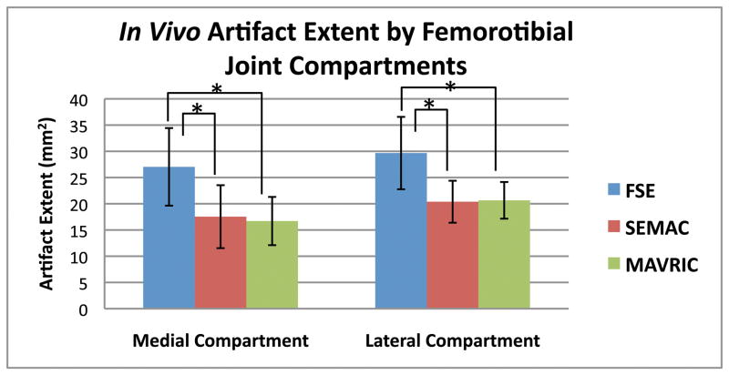
Figure 4.
Artifact extent measured on MR images of volunteers with TKRs showed that SEMAC and MAVRIC had significantly less artifact than conventional FSE images on all metal joint compartments of the knee (p < 0.0001). Correction of artifact is statistically similar between SEMAC and MAVRIC (p = 0.58). (* denotes statistical significance, p < 0.0083)

