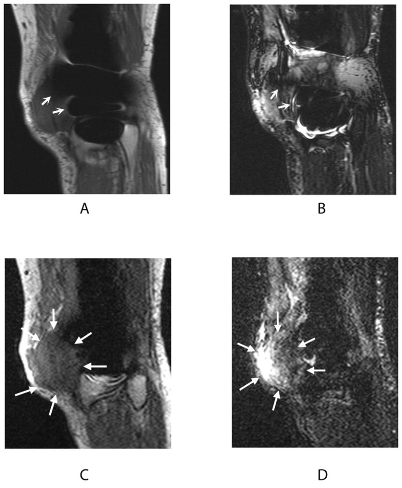
Figure 6.

Signal loss in 2D-FSE PD (a) and 2D-FSE IR (b) images (curved arrows) obscures the full extent of recurrent osteosarcoma visible on PD-SEMAC (c) and IR-SEMAC (d) images (wedge-shaped arrows). This finding on SEMAC images led to computed tomography (CT) scanning for lung metastases, confirmed at biopsy.
