
Figure 4.
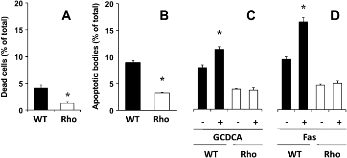
Proportion of dead cells (A) and hypodiploid bodies (B) determined with flow cytometry after staining with propidium iodide in wild-type (WT) and mtDNA-depleted (Rho) Hepa 1–6 mouse hepatoma cells. Effect of incubation with 100 µM glycochenodeoxycholic acid (GCDCA) (C) and 2 μg·mL−1 Fas antibody (Jo2) (D) for 24 h on the proportion of apoptotic bodies. Values are mean ± SEM from five cultures measured in triplicate for each data point. *P < 0.05, significant differences between WT and Rho cells.