
Figure 6.
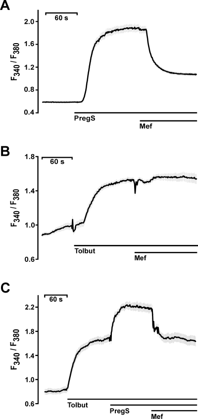
Effects of mefenamic acid on primary mouse pancreatic β-cells. Changes in [Ca2+]i are depicted by the fluorescence ratios F340/F380 of Fura-2-loaded primary mouse pancreatic islet cells. (A) Measurement of [Ca2+]i after stimulation with 35 µM pregnenolone sulphate and block with 30 µM mefenamic acid. (B) Mefenamic acid (30 µM) had no effect on [Ca2+]i increased by 100 µM tolbutamide (Tolbut). (C) Tolbutamide (100 µM) increased the [Ca2+]i in mouse β-cells. The addition of 35 µM pregnenolone sulphate further enhanced [Ca2+]i, an effect reversed by the application of mefenamic acid. The black lines indicate mean values from at least four independent experiments with at least 20 cells each. The shaded areas depict the SEM for each data point.