
Figure 2.
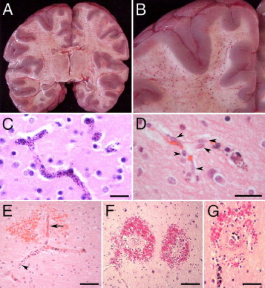
A and B: Coronal sections through the cerebral hemispheres of a CM2 patient. A: Numerous petechial hemorrhages are distributed throughout the white matter. The lateral and third ventricles are compressed because of edema. B: Close-up view of hemorrhages in the white matter. The gray matter is largely spared. C through G: Vascular changes in CM2 patients. C: The lumen of all small vessels in the field is distended by sequestered iRBCs. D: Some microvascular endothelial cells are hypertrophic and display large vesicular nuclei (arrowheads). E: A small branching vessel in the white matter contains iRBCs and hemozoin. One of the branches (arrow) is occluded by thrombus, is partially denuded of endothelial cells, and is associated with an RH. In the adjacent branch with iRBC sequestration, hypertrophic endothelial cells display large nuclei (arrowhead). F and G: RHs in the cerebral white matter correspond to petechial hemorrhages in A and B. G: Small, often thrombosed, ruptured capillaries containing iRBCs are immediately surrounded by a zone of necrosis that, in turn, is surrounded by a ring of extravasated RBCs, a few white blood cells, and extraerythrocytic pigment granules. C through G: H&E staining. Scale bars: 25 μm (C and D); 50 μm (E and G); 100 μm (F).