
Figure 4.
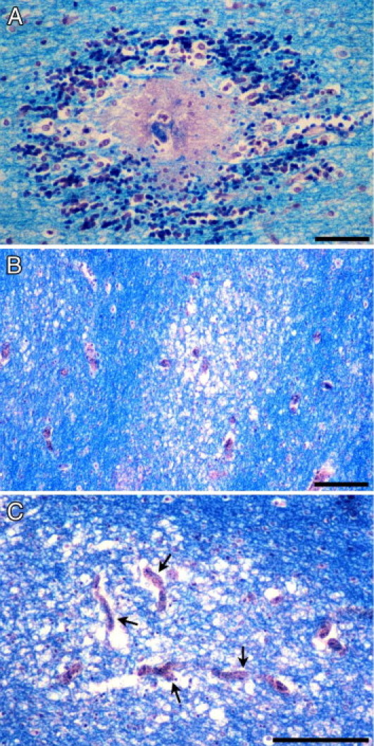
Myelin damage in pediatric CM. A: Myelin pallor and fragmentation has occurred in association with an RH surrounding a thrombosed and disrupted parasitized microvessel in the cerebral white matter. B: Irregular, ill-defined, and variably sized foci of diffuse myelin vacuolation and pallor unassociated with RHs typically occur in the CM2 and, to a lesser extent, CM1 groups. C: Areas of diffuse myelin damage encompass several distended microvessels filled with iRBCs (arrows). Luxol fast blue/H&E staining. Scale bars: 50 μm (A); 100 μm (B and C).