
Figure 9.
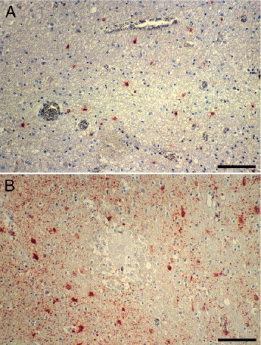
Reactive gliosis in CM detected by glial fibrillary acidic protein IHC. A: Mild diffuse gliosis in and around an area of myelin pallor and vacuolation and prominent iRBC sequestration. B: Diffuse gliosis in the neuropil around an RH. Scale bar = 100 μm (A and B).