
Figure 7.
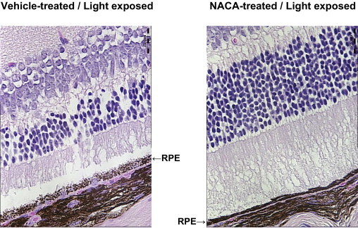
Maintenance of RPE cell integrity in vivo with NACA. Confocal microscopy (×630) obtained 6 days after light exposure. RPE cell disruption as induced by light exposure was significantly prevented with NACA pretreatment compared with vehicle alone. Representative images of H&E staining for retinal sections at the same location 400 μm superior to the optic nerve head demonstrating prevention of RPE disruption with NACA (right) compared with vehicle alone (left). Scale bars = 25 μm.