Figure 5.
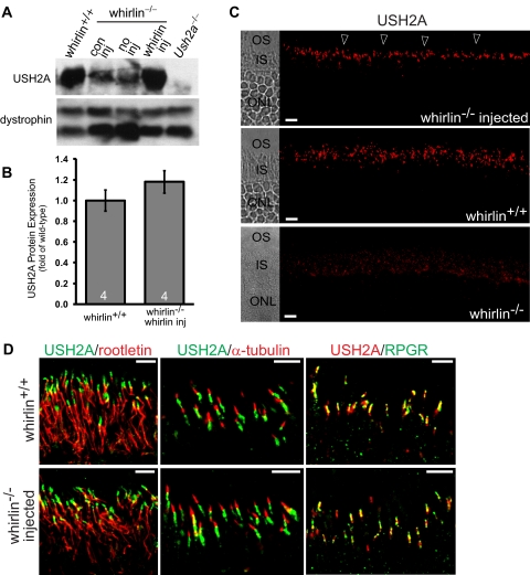
Restoration of USH2A expression in whirlin−/− retinas injected with AAV-whirlin. (A) A representative Western blot showing USH2A expression in the wild-type retina (whirlin+/+) and the whirlin−/− retina with different treatments: con inj, control subretinal injection with AAV-zsGreen, 10 weeks post injection; no inj, no injection; whirlin inj, subretinal injection with AAV-whirlin, 10 weeks post injection. The Ush2a knockout retina (Ush2a−/−) was used as a negative control. The dystrophin signal on the same blot (two bands) was used as a sample loading control. (B) Quantification of USH2A signal intensities on the Western blot from wild-type retinas with no subretinal injection and whirlin−/− retinas at 10 weeks post injection of AAV-whirlin. There is no statistically significant difference between the two groups. The number of mice measured in each group is indicated at the bottom of the bars. The error bar represents the SEM. (C) Immunostaining of USH2A in the whirlin−/− retina at 8 weeks post injection of AAV-whirlin (top panel, arrowheads), the wild-type retina (middle panel), and the whirlin−/− retina without any injection (bottom panel). The corresponding differential interference contrast images are shown on the left. OS, outer segment; IS, inner segment; ONL, outer nuclear layer. Scale bars, 10 μm. (D) Confocal images of USH2A double-stained with rootletin (left), acetylated α-tubulin (middle), and RPGR (right) in the wild-type retina (whirlin+/+, upper panels) and the whirlin−/− retina at six months post injection of AAV-whirlin (lower panels). These signal patterns strongly suggest that USH2A is localized at the PMC in the whirlin−/− retina injected with AAV-whirlin. Scale bars, 5 μm.

