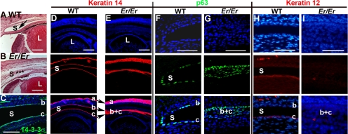
Figure 1.
Corneal defects in Er/Er mice at E18.5. (A, B) H&E staining of sagittal sections of WT and Er/Er corneas from E18.5 embryos. Arrow: the conjunctival sac (S) in the WT eye (A); (✶) the unseparated conjunctival sac in the Er/Er eye (B). (C) Immunostaining for14-3-3σ was evident on both conjunctival (b) and corneal (c) epithelial cells. (D, E) K14 immunostaining (red) was restricted to basal cells of epidermal (a), conjunctival (b), and corneal (c) epithelia in the WT control (D), but expanded in the skin epidermal layer (a), and the conjunctival sac failed to separate into conjunctival and cornea epithelia (b, c) in the Er/Er mice (E). (F, G) p63 (green) was normally restricted to the conjunctival basal progenitor cells, which line the single layer of conjunctival and corneal epithelial cells (F), but the conjunctival sac in the Er/Er mice was not completely formed, and it filled with the expanded p63-positive cells (G). The corneal epithelial marker, K12 was labeled red in the WT (H), but not in the Er/Er (I) corneas. Top: nuclei counterstained with DAPI; middle: antibody staining; bottom: merged images. L, lens. Small letters mark skin (a), conjunctival (b), and corneal (c) epithelia. (b, c) The unseparated conjunctival and corneal epithelial fusion. Scale bars: (A–E) 100 μm; (F–I) 50 μm.

