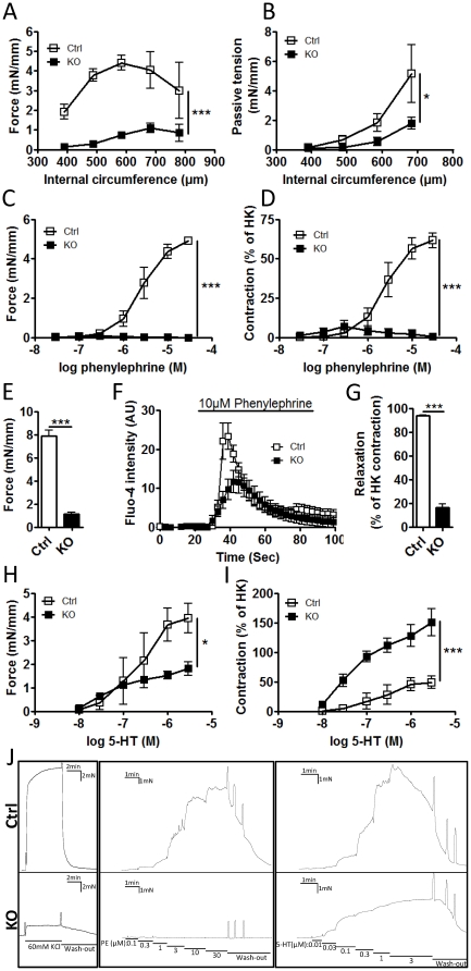
Figure 3. Severe impairment of contractile function in SM-Dicer KO arteries.
(A) Contractile function of control and SM Dicer KO arteries was analyzed in a wire myograph, 10 weeks post Tamoxifen treatment. The active and passive circumference-tension relationships were analyzed in control (Ctrl) and SM-Dicer KO (KO) small mesenteric arteries. Active force in response to 80 mM KCl (HK) at different circumferences is summarized in A. In B, summarized data of the passive tension in nominally calcium free condition is shown. (C) Contractile responses to phenylephrine were measured in saphenous arteries and shown as absolute force or (D) force in relation to HK-induced responses. (E) Force in response to KCl in saphenous arteries is shown separately. (F) Relative phenylephrine-induced calcium influx in isolated aortic SMCs from Ctrl and KO mice were measured using Fluo-4 calcium indicator. (G) Represents summarized data of the remaining contractile tone following 1 min washout of the HK-induced contraction. Contractile responses to serotonin (5-HT) were measured in saphenous arteries and shown as absolute force (H) or force in relation to KCl-induced responses (I) (n = 3–4 mice for all experiments). An original trace recording from a representative experiment on saphenous artery is shown in J.

