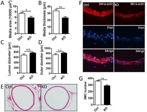
Figure 4. Morphological changes in the aorta of SM-Dicer KO mice.
(A) Perfusion fixed and paraffin embedded sections of the aorta of control (Ctrl) and SM-Dicer KO (KO) mice were analyzed for morphological changes 10 weeks post tamoxifen treatment. Summarized data from 4–5 animals in shown in A-D. (E) Representative image of H&E stained Ctrl and KO aorta. (F) Representative images of Ctrl and KO aortas stained for SM-α-actin (red) and Dapi (blue). (G) Summarized data of SMC number in Ctrl and SM-Dicer KO aorta.

