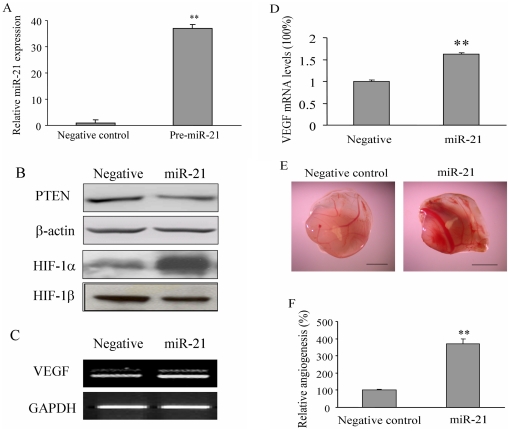
Figure 1. MicroRNA miR-21 overexpression increased HIF-1α and VEGF expression and induced tumor angiogenesis.
(A) Human prostate cancer cells DU145 were transfected with pre-miRNA negative control or pre-miR-21 at 25 nM. After the transfection for 36 h, cells were collected and subjected to qRT-PCR for miR-21 expression. (B) DU145 cells were treated as in A, the total protein was extracted and subjected to Western blotting assay. (C–D) DU145 cells were collected after the transfection for 36 h. Total RNAs were extracted and analyzed for VEGF and GAPDH mRNA expression by RT-PCR analysis (C), and by real-time RT-PCR (D). (E) DU145 cells were transfected as above. After 24 h, cells (2×10⁶cells, 15 µl) were mixed with 15 µl Matrigel. The cell mixture was implanted onto the CAM of a 9-day old chicken embryo. After 4 days of implantation, the CAM was cut off, and the amount of blood vessels on the CAM induced by the DU145 plugs was determined from eight different replicated experiments. Representative plugs from negative control and miR-21 treatment groups were shown. Scale, 2 mm. (F) The number of blood vessels were counted from replicate experiments, and normalized to that of the negative control group as relative angiogenesis. The data are mean±SD from 6 replicates. ** indicates the significant increase when compared to the pre-miRNA negative control (p<0.01).

