
Fig. 4. Maternally-derived IL-6 activates the JAK/STAT3 pathway in the fetal placental compartment in response to MIA.
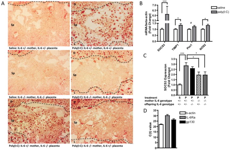
A. Representative sections of the spongiotrophoblast layer of the placenta (with decidua along the top edge and labyrinth along the bottom edge of each image) show positive pSTAT3 staining when placentas are isolated from poly(I:C)-injected mothers. Eliminating the contribution of fetally-derived IL-6 has no effect on pSTAT3 staining, while eliminating the contribution of maternally-derived IL-6 completely abrogates pSTAT3 staining in the spongiotrophoblast. [n=5-7 placentas per treatment group per genotype pair (pooled from 3-5 independent litters)]. Dotted lines = boundary between decidua and spongiotrophoblast layers (upper) and boundary between spongiotrophoblast and labyrinth (lower); D=decidua, Sp=spongiotrophoblast, L=labyrinth; B. MIA-induced JAK/STAT3 activation is accompanied by increased expression of the downstream acute phase genes, SOCS3, TIMP1, Pim1 and NOS2, in poly(I:C) placentas [n=9 placentas per treatment group (pooled from 3 independent litters); ***p < 0.0001, *p < 0.05, # p = 0.07]. C. Eliminating the contribution of maternally-derived IL-6 reduces placental SOCS3 induction [n=9 placentas per treatment group per genotype pair (pooled from 3 independent litters); ***p < 0.0001, *p < 0.05]. D. Murine spongiotrophoblast cells express IL-6Rα and gp130 mRNA, indicating that they may respond directly to placental IL-6 activation. [n=3 saline, 3 poly(I:C} placentas; merged].