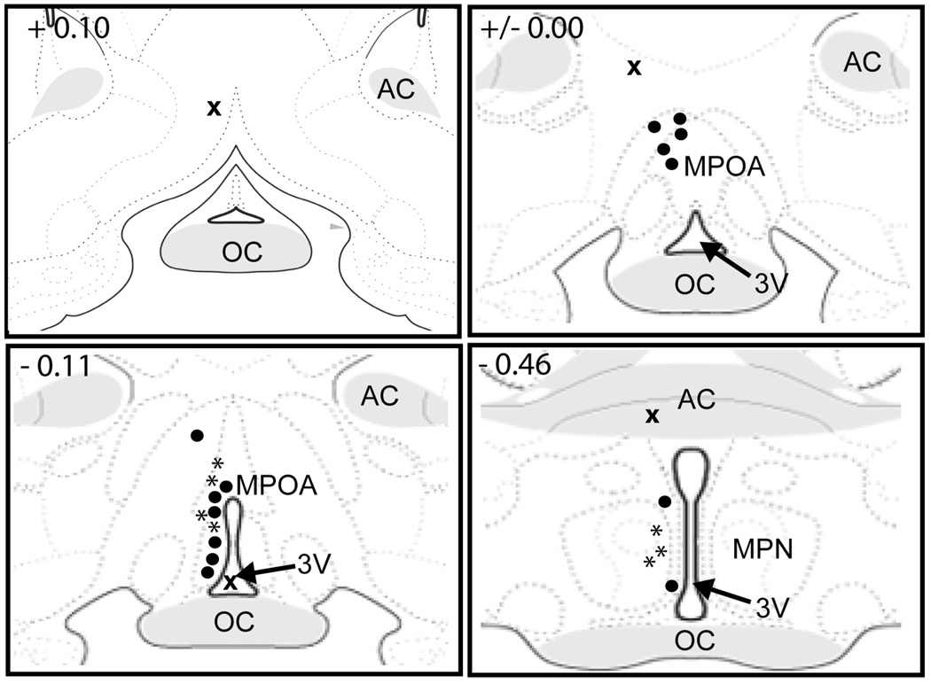
Fig. 3. Representative histology showing cannula placements for intra-MPOA injections of an OTA.
Dark circles represent placements within the MPOA (n=14); an “x” represents a placement outside of the MPOA (n=4); stars represent placements in the MPOA for additional animals that were used in the locomotor experiment (n=7). MPOA, medial preoptic area; MPN, medial preoptic nucleus; AC, anterior commissure; OC, optic chiasm; 3V, 3rd ventricle. Images adapted and modified from Swanson (2003), with permission from Elsevier.

