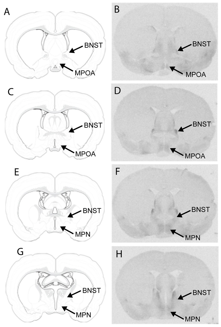
Fig. 4. Autoradiograms showing oxytocin receptor binding in the MPOA.
rostal MPOA 1 (B), rostral MPOA 2 (D), MPN (F), and caudal MPN (H). Illustrations adapted from Swanson (2003), with permission from Elsevier, showing: rostral MPOA 1 (A), rostral MPOA 2 (C), MPN (E), and caudal MPN (G). MPOA, medial preoptic area; MPN, medial preoptic nucleus; BNST, bed nucleus of the stria terminalis.

