
Figure 4.
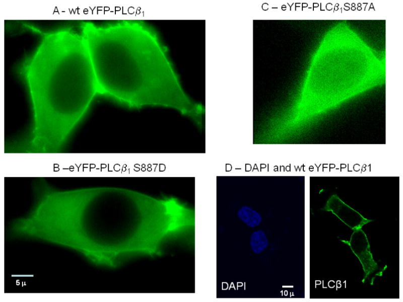
Representative images of HEK293 cells transfected with eYFP-wtPLCβ1 (A), eYFP-S887D (B) and eYFP-S887D where the cells were imaged 48 hours after transfection and viewed through a YFP filter and a 60× objective (see [6] for details). The images shown are from a 0.2μ slice in the middle of the cell so that the nuclear region can clearly be seen. Note that only the S887A mutant had a significant nuclear population. D – A side-by-side comparison of fixed HEK293 cells stained with DAPI to view the nuclei (left panel) of cells expressing eYFP-wtPLCβ1 (right panel),