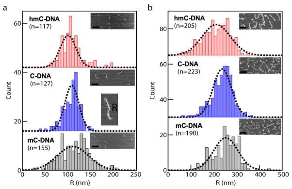
Figure 6.
AFM analysis of DNA with modified cytosines. (a) End-to-end distances R measured by tapping mode AFM of 410 bp fragments immobilized on mica by incubation with a solution of DNA containing 2 mM Tris and 1 mM Mg2+. Insets show representative AFM images for each of the samples (scale bar = 200 nm). The number of molecules n in each distribution is indicated. (b) Similar analysis as in (a) for an 1100 bp DNA fragment. For both DNA lengths, R follows hmC-DNA<C-DNA<mC-DNA.

