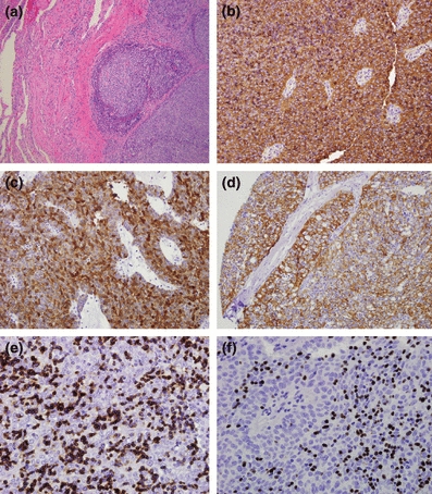
Figure 4.

Type B3 thymoma. (a) Atypical thymic cell proliferation with pushing borders to adjacent lung tissue (H&E staining, 20×) (b) CD57 diffuse strong staining (10×); (c) Androgen receptor diffuse strong cytoplasmic staining (10×); (d) Cytokeratin 5/6 and diffuse moderate to strong staining (10×); (e) CD1a stating immature lymphocytes (10×); (f) TdT staining immature lymphocytes (10×).
