
Figure 1.
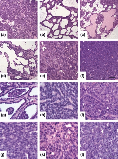
(a–f) Assessment of tumour morphology in H&E-stained MMTV-PyVmT tumours. (a–d) well-defined, non-invasive lesions characteristic of early grade tumours. (a) acinar; tumour comprised of small glandular clusters around small lumens. (b) cribiform; nests of cells forming large lumens. (c) glandular; tumour composed of glands, with some glands dilated by protein-rich secretion. (d) papillary; tumour forms epithelial-lined projections with a fibro-vascular core. (e–f) poorly defined, invasive lesions characteristic of mid – late grade tumours. (e) acinar pattern with focal invasion; boundaries between clusters of cells become unclear. (f) solid; tumour is composed of solid sheets of cells, with no glandular differentiation. Scale bar in (f) represents 100 μm. (g–l) Assessment of nuclear grade in H&E stained MMTV-PyVmT tumours. (g, h) grade 1 (low); heterochromatic nuclei relatively uniform in size and shape with a slight increase in nuclear/cytoplasmic ratio as compared to normal ductal cells. (i, j) grade 2 (intermediate); an increasing number of cells have euchromatic nuclei (nucleoli may or may not be visible). Moderate heterogeneity in nuclear size and shape with a moderate increase in nuclear/cytoplasmic ratio. (k, l) grade 3 (high); nuclear pleomorphism, with a marked increase in nuclear/cytoplasmic ratio and clearly visible nucleoli. Scale bar in (l) represents 30 μm.