Figure 1.
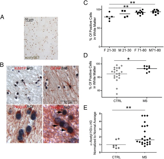
Histone H3 acetylation in the frontal cortex of MS and normal brains. A, Immunocytochemical analysis of histone H3 acetylation of white matter in the frontal lobes of control subjects, without neurological disease. B, Double stainings were performed with antibodies specific for acetyl-H3 (AcH3) (black) and either NogoA (red) to identify oligodendrocytes, KiM1P to identify microglia, and GFAP to label astrocytes. Examples of an AcH3-negative microglial cell, an AcH3-positive astrocyte, and AcH3-positive (arrows) and -negative oligodendrocytes are shown. C, Quantification of nuclei immunoreactive for acetylated histone H3 in the white matter of the frontal lobes of young (21–30 years) and old (71–80 years) male (squares) and female (circles) subjects. D, Quantification of total acetyl-H3+ nuclei in the frontal cortex of non-neurological (CTRL) and MS patients demonstrating a significantly increased percentage of acetyl-H3+ nuclei in chronic MS brains (Mann–Whitney test; *p < 0.05). E, Scatterplot of the amount of acetylated H3 protein levels isolated from CTRL and MS patients. Proteins were isolated from frozen frontal lobes perilesional and NAWM of control and MS patients and processed by slot blot using antibodies against acetyl-H3 and total H3 to measure global levels of acetylation on the tail of histone H3 (two-tailed t test; *p < 0.05; **p < 0.01).

