
Figure 2. βPAT-3 localizes normally in muscle cells of rescued animals.
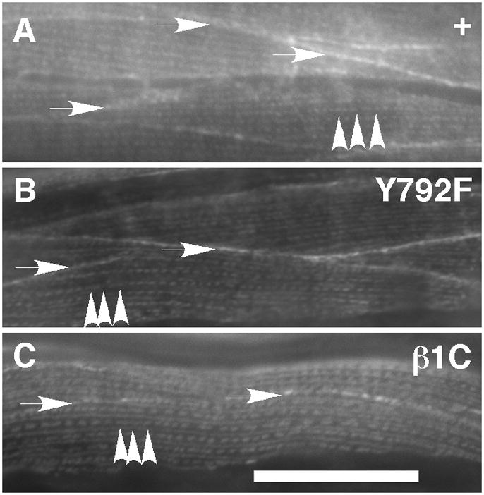
(A) βPAT-3 in rescued lines was detected by immunofluorescence with MH25 antibodies. Typical dense body (arrowheads) and cell-to-cell contacts (arrows) were observed in the muscle cells. The staining pattern was similar to N2 (data not shown). This typical MH25 staining pattern was observed in other transgenic lines such as (B) βpat-3(Y792F) and (C) βpat-3(β1C). Scale bars= 40 μm.