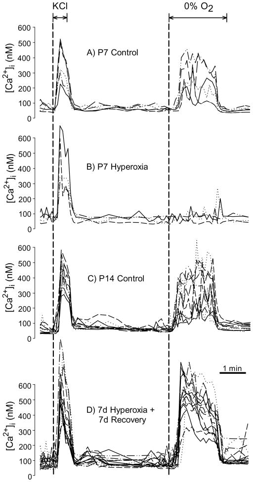
Fig. 3.
Representative glomus cell intracellular calcium ([Ca2+]i) responses to hypoxia for rats reared in 21% O2 (Control) or 60% O2 (Hyperoxia) through P7 (panels A, B) and after 7 d recovery in normoxia (i.e., P14; panels C, D). Each graph panel contains data from several glomus cells plated on a single coverslip, with each line representing serial [Ca2+]i measurements for a single cell during exposure for 30 s to 20mM KCl followed by a 2 min hypoxia challenge (0% O2). Note that glomus cells from P7 Hyperoxia rats remained responsive to KCl (a nonspecific depolarizing stimulus) while the response to hypoxia was markedly reduced.

