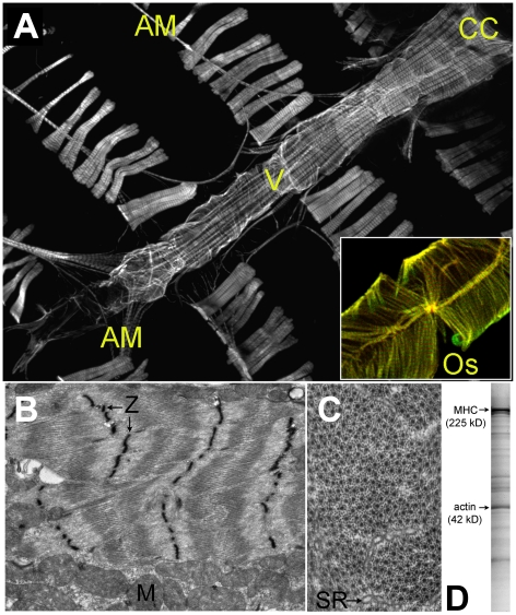
Figure 1. The Cardiac Tube of Drosophila melanogaster.
Panel A. TRITC-Phalloidin labeled wild-type Drosophila heart tube and associated structures (10× magnification). CC = conical chamber; AM = alary muscle; v = internal valve; Os = ostia in flow tract. Inset: luminal surface of TRITC-Phalloidin-labeled myosin-GFP-expressing heart (20× magnification). Ostia inflow tracts and the striated alternating myosin and actin myofilament bands are clearly resolved. Panel B. Electron micrograph of a longitudinal section through the conical chamber reveals the contractile myofibrils and mitochondria (M)(3,800×). Densely stained Z-bands (Z) demarcate individual sarcomeres and bisect the I-bands. Centrally-located A-bands are also apparent. Panel C. Cross-section through cardiac myofibrils of the conical chamber (10,500×). Individual thick filaments are surrounded by 9–11 thin filaments. Regions of sarcoplasmic reticulum (SR) can also be resolved. Panel D. 10% Coomassie-stained polyacrylamide gel from 30 Drosophila heart tubes. Sarcomeric myosin heavy chain (MHC) and actin are highlighted for reference.

