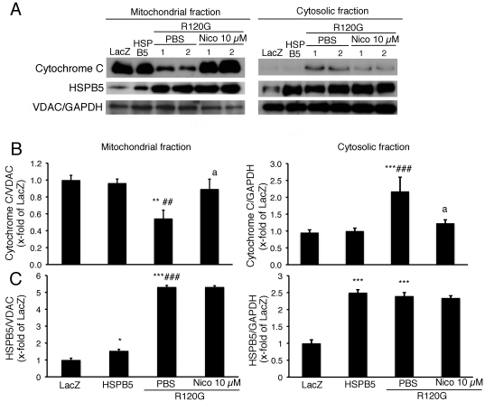
Figure 2. Cytochrome c levels of mitochondrial fraction isolated from the cardiomyocytes expressing HSPB5 R120G (R120G) with or without Nico treatment.
(A) Typical pictures of Western blot analysis. Quantitative analysis of cytochrome c (B) and HSPB5 (C) in mitochondrial and cytosolic fractions isolated from the cardiomyocytes. GAPDH was used as a loading control in the cytosolic fraction and the voltage-dependent anion channel (VDAC) was used in mitochondrial fraction. *p<0.05, ** p<0.01, ***p<0.001 vs. the cardiomyocytes infected with LacZ (LacZ), ##p<0.01, ###p<0.001 vs. the cardiomyocytes infected with wild-type HSPB5 (HSPB5), ap<0.05 vs. cardiomyocytes infected with R120G treated with PBS. (n = 6).

