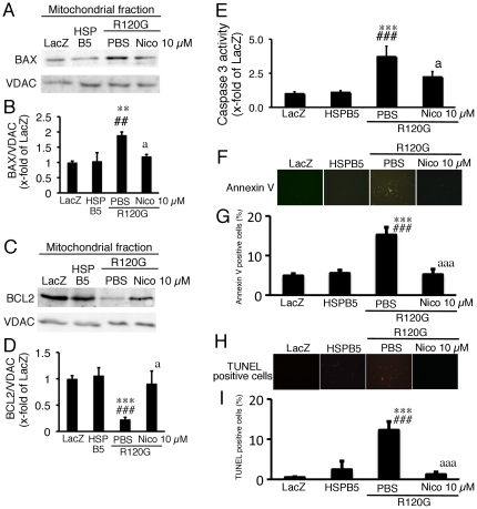
Figure 3. Western blot analysis of apoptosis related proteins, BAX and BCL2, and apoptotic cell death in the cardiomyocytes expressing HSPB5 R120G (R120G) with or without Nico treatment.
(A) Typical pictures of Western blot analysis. (B) Quantitative analysis of BAX in mitochondrial fraction isolated from the cardiomyocytes. (C) Typical pictures of Western blot analysis. (D) Quantitative analysis of BCL2 in mitochondrial fraction isolated from the cardiomyocytes. (E) Caspase 3 activity in the cardiomyocytes. (F) Typical pictures of annexin V-positive cardiomyocytes. (G) Quantitative analysis of annexin V-positive cardiomyocytes. (H) Typical pictures of TUNEL-positive cardiomyocytes. (I) Quantitative analysis of TUNEL-positive cardiomyocytes. ** p<0.01, ***p<0.001 vs. the cardiomyocytes infected with LacZ (LacZ), ##p<0.01, ###p<0.001 vs. the cardiomyocytes infected with wild-type HSPB5 (HSPB5), ap<0.05, aaap<0.001 vs. cardiomyocytes infected with R120G treated with PBS. (n = 6).

